Cellular Maestros: ER and GERL in Symphonic Synthesis and Precision Delivery
Golgi Apparatus / ER-Golgi Intermediate Compartment (GERL) System:
Question: What is the literal meaning of endoplasmic reticulum?
Answer: The literal meaning of "Endoplasmic Reticulum" is derived from its Latin and Greek roots. The Greek prefix "endo" translates to "inside" or "within," while "plasma" refers to a form of matter. The Latin term "Reticulum" means "little net" or "network." Combining these elements, the
The literal interpretation of Endoplasmic Reticulum is "within the cell matter network" or "inside the cell's net-like structure." This name reflects the appearance of the organelle, which consists of a complex network of membrane-enclosed tubules and sacs within the cell.
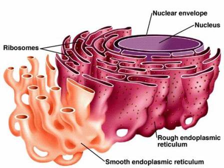
Endoplasmic Reticulum Structure:
1. Rough Endoplasmic Reticulum (RER):
- The RER is characterized by the presence of ribosomes attached to its cytoplasmic surface, giving it a "rough" appearance under a microscope.
- These ribosomes are involved in protein synthesis and play a crucial role in the production of membrane and secretory proteins.
- The RER is often found adjacent to the nucleus, reflecting its role in the synthesis of proteins destined for various cellular compartments.
2. Smooth Endoplasmic Reticulum (SER):
- The SER lacks ribosomes on its cytoplasmic surface, giving it a "smooth" appearance.
- It is involved in various metabolic processes, including lipid synthesis, detoxification reactions, and calcium ion storage.
- The SER is often more prevalent in cells with specialized functions, such as liver cells that participate in detoxification processes.
3. Interconnected Membrane Network:
- Both the RER and SER are part of the same membrane system, forming a highly interconnected network of tubules and sacs throughout the cell.
- This network allows for the efficient transport of materials within the cell and facilitates communication between different cellular compartments.
4. Continuous with the Nuclear Envelope:
- The ER is continuous with the outer membrane of the nuclear envelope, emphasizing its role in membrane synthesis and maintaining cellular structure.
5. Dynamic Nature:
- The structure of the endoplasmic reticulum is dynamic, with membranes constantly undergoing processes like fusion, fission, and rearrangement to meet the changing needs of the cell.
Endoplasmic Reticulum Composition:
1. Membranes:
- The endoplasmic reticulum primarily consists of a complex network of membranes.
- This membrane system forms a dynamic and interconnected structure composed of tubules and flattened sacs known as cisternae.
2. Tubules and Cisternae:
- Tubules: These are narrow, elongated structures that make up the branching network of the ER. They are involved in the transport of materials within the cell.
- Cisternae: These are flattened, disk-like sacs that are interconnected with the tubules. Cisternae provide a larger surface area for various cellular processes, such as protein and lipid synthesis.
3. Ribosomes:
- In the Rough Endoplasmic Reticulum (RER), ribosomes are attached to the cytoplasmic surface of the membranes, particularly to the outer surface of the cisternae.
- Ribosomes are responsible for synthesizing proteins that are either incorporated into the membranes or secreted from the cell.
4. Enzymes:
- The endoplasmic reticulum contains various enzymes associated with the membranes. These enzymes play crucial roles in processes such as protein folding, modification, and quality control.
5. Lipids:
- Lipids, including phospholipids, steroids, and triglycerides, are synthesized in the Smooth Endoplasmic Reticulum (SER), contributing to the structure and function of cellular membranes.
6. Calcium Ion Stores:
- The SER serves as a storage site for calcium ions within vacuoles, contributing to calcium homeostasis within the cell.
7. Vacuoles:
- Vacuoles within the endoplasmic reticulum serve as storage compartments for various substances, including ions and molecules.
- Vacuoles play a role in maintaining cellular homeostasis by storing and releasing substances as needed.
8. Size in Angstroms:
- The size of the components within the endoplasmic reticulum varies. The diameter of ER tubules can range from approximately 50 to 100 Angstroms.
- The cisternae have larger dimensions, with their width ranging from around 500 to 1000 Angstroms.
- Specific sizes of ER vacuoles can vary depending on their content and function, with dimensions ranging from tens to hundreds of Angstroms.
Question: Why and How Does Rough Endoplasmic Reticulum (RER) Change into Smooth Endoplasmic Reticulum (SER) or Vice Versa?
Answer: The transformation of Rough Endoplasmic Reticulum (RER) into Smooth Endoplasmic Reticulum (SER) or vice versa is a dynamic process influenced by the cellular needs and metabolic demands of the cell. The transition involves various factors and mechanisms that contribute to the adaptability of the endoplasmic reticulum (ER) to changing conditions.
Factors Influencing the Transformation:
-
Protein Synthesis vs. Lipid Synthesis
- RER, involved in protein synthesis, undergoes changes in response to variations in the demand for protein production.
- Reduction or loss of ribosomes on the RER surface can occur when the cell requires a shift towards lipid synthesis or other functions associated with the SER.
-
Dynamic Membrane Adjustments:
- The ER's dynamic nature allows for continuous adjustments in membrane structure through processes like fusion, fission, and rearrangement.
- These dynamic membrane adjustments contribute to the flexibility of the ER to adapt to changing cellular demands.
-
Cellular Signaling and Regulation:
- Cellular signals and regulatory mechanisms play a crucial role in maintaining the balance between RER and SER.
- Signaling pathways influenced by cellular conditions and external cues modulate the expression of proteins involved in membrane synthesis, protein synthesis, and lipid metabolism.
-
Cell Differentiation and Specialization:
- Cellular differentiation leads to changes in the ER composition to meet specific functional requirements.
- Cells with a high demand for lipid synthesis may exhibit a more prominent smooth endoplasmic reticulum.
-
Cellular Stress and Responses:
- Environmental stress or specific cellular conditions impact the balance between RER and SER.
- Exposure to toxins, for example, may induce the synthesis of detoxification enzymes associated with the smooth endoplasmic reticulum.
Question: What is Sarcoplasmic Reticulum (SR), and How Does it Differ from Endoplasmic Reticulum (ER)?
Answer: The sarcoplasmic reticulum (SR) is a specialized form of endoplasmic reticulum (ER) found in muscle cells, specifically in the sarcoplasm, the cytoplasm of muscle cells. While both the sarcoplasmic reticulum and endoplasmic reticulum share some similarities, they also have distinct structural and functional differences.
Sarcoplasmic Reticulum (SR):
-
Location:
- The SR is located in muscle cells, particularly in close association with the myofibrils within the sarcoplasm.
-
Function:
- It primarily regulates calcium ion levels within muscle cells, storing and releasing calcium ions during muscle contraction and relaxation.
-
Structure:
- The SR has a specialized structure, forming a network of tubules and vesicles around the myofibrils to facilitate the controlled release of calcium ions.
-
Relationship with Muscle Contraction:
- The release of calcium ions from the SR triggers muscle contraction by binding to troponin, a regulatory protein involved in the sliding of actin and myosin filaments.
Endoplasmic Reticulum (ER):
-
General Location:
- The endoplasmic reticulum is a network of membranes found in the cytoplasm of eukaryotic cells, including non-muscle cells.
-
Functions:
- In non-muscle cells, the ER is involved in various cellular processes, such as protein synthesis, lipid metabolism, and detoxification.
-
Types of ER:
- There are two main types of endoplasmic reticulum: the rough endoplasmic reticulum (RER), involved in protein synthesis, and the smooth endoplasmic reticulum (SER), involved in lipid synthesis and detoxification.
-
Structural Differences:
- The ER has a more diverse structure, encompassing both rough and smooth regions with various functions depending on the cell type.
Question: Which Cells Do Not Contain a Well-Developed Endoplasmic Reticulum (ER) in Humans?
Answer: There are three main types of cells in the human body that lack a well-developed endoplasmic reticulum (ER):
-
Mature Red Blood Cells (Erythrocytes):
- Mature red blood cells lose their nucleus and most organelles, including the endoplasmic reticulum, during development.
- This adaptation allows red blood cells to have more space for hemoglobin, facilitating their primary function of oxygen transport.
-
Platelets (Thrombocytes):
- Platelets, which are fragments of large bone marrow cells called megakaryocytes, also lack a well-developed endoplasmic reticulum.
- Platelets are involved in blood clotting, and their structure is adapted to their specific functions, which do not heavily rely on extensive protein and lipid synthesis facilitated by the ER.
-
Mature Sex Cells (Sperm and Eggs):
- Mature sperm cells in males and ova (eggs) in females also lack a well-developed endoplasmic reticulum.
- During the maturation process of sex cells (spermatogenesis in males and oogenesis in females), there is a reduction in cellular organelles, including the endoplasmic reticulum, to streamline the cells for their specific functions—sperm motility and fertilization for sperm, and fertilization and subsequent embryonic development for eggs.
Question: What is the Role of Endoplasmic Reticulum (ER) in Co-Translational Transport?
Answer: The endoplasmic reticulum (ER) plays a pivotal role in co-translational transport, a process closely linked to protein synthesis. Several key components of this process take place within the ER, emphasizing its significance in the maturation and transport of proteins.
Co-Translational Transport:
-
Initiation of Protein Synthesis:
- Ribosomes, which are responsible for protein synthesis, attach to the rough endoplasmic reticulum (RER) during the initiation phase of translation.
-
Protein Synthesis on the ER Surface:
- The ribosomes on the RER surface facilitate the co-translational synthesis of proteins that are destined for secretion, membrane incorporation, or endomembrane system components.
-
Signal Recognition Particle (SRP):
- During translation, a signal recognition particle (SRP) recognizes the signal peptide emerging from the ribosome.
- The SRP binds to the ribosome, temporarily halting translation.
-
Targeting the ER:
- The ribosome-SRP complex docks onto the ER membrane, where the translocon (a protein-conducting channel) facilitates the translocation of the growing polypeptide chain into the ER lumen.
-
Co-Translational Translocation:
- Co-translational transport involves the simultaneous synthesis and translocation of a polypeptide into the ER, ensuring proper folding and modification during translation.
-
Protein Folding and Modification:
- Once inside the ER lumen, proteins undergo folding and post-translational modifications, including glycosylation and disulfide bond formation.
-
Quality Control Mechanisms:
- The ER monitors the quality of newly synthesized proteins. Misfolded or aberrant proteins may be targeted for degradation through a process known as ER-associated degradation (ERAD).
-
Export of proteins:
- Properly folded and modified proteins are transported from the ER to their final destinations, such as the Golgi apparatus, vesicles, or the cell membrane.
Question: What are the functions of smooth endoplasmic reticulum (SER) and rough endoplasmic reticulum (RER)?
Answer: The endoplasmic reticulum (ER) is a crucial organelle with two distinct regions: the Smooth Endoplasmic Reticulum (SER) and the Rough Endoplasmic Reticulum (RER). Each region has specialized functions contributing to diverse cellular processes.
Smooth Endoplasmic Reticulum (SER):
-
Lipid Synthesis:
- The primary function of the SER is lipid synthesis, including the production of phospholipids and steroids.
- It plays a key role in the synthesis of lipids needed for cellular membranes and various cellular processes.
-
Detoxification:
- The SER is involved in the detoxification of drugs and poisons within the cell.
- Enzymes in the SER contribute to the breakdown of potentially harmful substances, enhancing cellular defense mechanisms.
-
Calcium Ion Storage:
- SER functions as a calcium ion reservoir, participating in the regulation of intracellular calcium levels.
- Stored calcium ions can be released when needed for signaling, muscle contraction, and the transmission of nerve impulses.
-
Transmission of Nerve Impulses:
- In certain cell types, the SER is involved in the transmission of nerve impulses by regulating calcium ion levels crucial for neuronal signaling.
-
Mechanical Support:
- The SER may provide mechanical support to the cell by synthesizing lipids that contribute to the structure and fluidity of cellular membranes.
Rough Endoplasmic Reticulum (RER):
-
Protein Synthesis:
- The primary function of the RER is protein synthesis.
- Ribosomes on the surface of the RER are responsible for the translation of mRNA into proteins, particularly those destined for secretion or integration into cellular membranes.
-
Membrane Synthesis:
- RER is involved in the synthesis of membrane proteins and proteins that are directed to various cellular compartments, including the endomembrane system.
-
Protein Folding and Modification:
- The RER provides a site for proper protein folding and post-translational modifications, such as glycosylation and disulfide bond formation.
-
Quality Control:
- The RER includes quality control mechanisms to ensure that newly synthesized proteins attain the correct conformation.
- Misfolded proteins may be targeted for degradation through ER-associated degradation (ERAD).
Golgi Apparatus/ER-Golgi Intermediate Compartment (GERL) System:
1. Structure:
- The Golgi Apparatus is a stack of flattened membrane-bound sacs called cisternae.
- The GERL (ER-Golgi Intermediate Compartment) is a region that connects the ER and the Golgi apparatus.
2. Functions:
- Protein Modification and Sorting: The Golgi Apparatus receives proteins from the ER, modifies them (e.g., glycosylation), and sorts them for delivery to specific cellular locations.
- Vesicle Formation: The Golgi apparatus forms vesicles that transport modified proteins to various destinations, including lysosomes, the plasma membrane, or other organelles.
- Polysaccharide Synthesis: It is involved in synthesizing certain polysaccharides.
3. Interconnectedness:
- The GERL acts as an intermediate compartment connecting the ER and the Golgi apparatus.
- It facilitates the transport of proteins from the ER to the Golgi apparatus for further processing and sorting.
Overall Connection:
- Proteins synthesized on the RER are transported to the Golgi apparatus through vesicles, where they undergo further modification, sorting, and packaging for various cellular functions.
1. Structure:
- The Golgi Apparatus is a stack of flattened membrane-bound sacs called cisternae.
- The GERL (ER-Golgi Intermediate Compartment) is a region that connects the ER and the Golgi Apparatus.
2. Functions:
- Protein Modification and Sorting: The Golgi Apparatus receives proteins from the ER, modifies them (e.g., glycosylation), and sorts them for delivery to specific cellular locations.
- Vesicle Formation: The Golgi apparatus forms vesicles that transport modified proteins to various destinations, including lysosomes, the plasma membrane, or other organelles.
- Polysaccharide Synthesis: It is involved in synthesizing certain polysaccharides.
3. Interconnectedness:
- The GERL acts as an intermediate compartment connecting the ER and the Golgi Apparatus.
- It facilitates the transport of proteins from the ER to the Golgi Apparatus for further processing and sorting.
Overall Connection:
- Proteins synthesized on the RER are transported to the Golgi Apparatus through vesicles, where they undergo further modification, sorting, and packaging for various cellular functions.

.jpg)


0 Comments