Exploring the Biochemical Composition of Charophytes: Insights into Cell Wall Components and Evolutionary Significance
Introduction to Charophyta:
Charophytes represent a group of green algae that exhibit advanced characteristics, often earning them the designation "macroscopic algae," as they resemble plants in appearance and, to some extent, in function. These unique organisms are distributed across all continents, excluding Antarctica. Their historical significance is rooted in the fact that they played a crucial role in the transition of plants from aquatic to terrestrial environments. Fossil evidence of charophytes dates back to the Silurian Period, around 443 million years ago, making them the closest living relatives to land plants.
Node:
Previously classified under the division Chlorophyta in the class Charophyceae, Charophytes have since been reclassified as a distinct division due to their distinctive features. These features include the differentiation of the plant body into nodes and internodes, as well as highly advanced oogamous sexual reproduction. The reproductive organs, such as the male Globule and the female Nucule, are specialized, and asexual reproduction via spore formation is absent.

https://www.inaturalist.org/
Distinctive Features of Charophyta:
-
Differentiation of the Plant Body:
- The plant body of charophytes is characterized by distinct nodes and internodes.
-
Highly Advanced Oogamous Reproduction:
- Charophytes employ a sophisticated oogamous method of sexual reproduction involving specialized reproductive organs.
-
Specialized Reproductive Organs:
- Male reproductive organs are represented by the Globule, while female reproductive organs are represented by the Nucule.
-
Absence of Asexual Reproduction by Spore Formation:
- Unlike some other algae, charophytes do not reproduce asexually through spore formation.
-
Freshwater Habitat:
- Charophytes are primarily restricted to freshwater habitats.
- further distinguishing them from other algae.
Characteristic Features:
-
Habitat:
- Members of Charophyta commonly grow in freshwater with muddy or sandy bottoms. They are also found in water flowing over limestone but are not present in saltwater.
-
Plant Body Organization:
- The plant body of Charophyta resembles that of Equisetum and Ephedra in its organization. It is differentiated into nodes and internodes, forming a jointed structure.
-
Branches and Growth:
- Branches of limited growth in Chara correspond to the leaves of higher plants.
-
Cell Wall Composition:
- The cell wall is mainly composed of cellulose, similar to plants and other green algae.
-
Flagella:
- They possess two flagella of the same length (isokont).
-
Chloroplasts and Pigments:
- Pigments are located in chloroplasts, which also contain a pyrenoid.
-
Reserve food material:
- The reserve food material is starch, composed of amylose and amylopectin.
-
Calcium Carbonate Precipitation:
- Some species of Charophyta (Chara and Nitella) have the ability to precipitate calcium carbonate from the water and cover themselves with a calcareous layer.
-
Reproduction:
- They reproduce through vegetative and sexual reproduction.
-
Asexual Reproduction:
- Asexual reproduction is not found.
-
Sexual Reproduction:
- Sexual reproduction is highly advanced and follows the oogamous type.
-
Diploid Structure:
- The zygote, or oospore, is the only diploid structure in their life cycle.
-
Position of Nucule and Globule:
- In Chara, the Nucule is present above the Globule, while in Nitella, it is reversed.
-
Monoecious Species:
- Monoecious species of Chara are protandrous.
Reproduction:
Vegetative Reproduction:
- By Tubers and Bulbils:
- Tubers are commonly formed on rhizoids or sometimes on buried nodes. They are starch-filled and may develop into a separate plant. Simple tubers may form when the globule divides.
- By Amylum or Starch Stars:
- The cells of some subterranean nodes become star-shaped and heavily laden with starch, known as amylum stars, which can develop into new plants.
- By Secondary Protonema:
- Protonema-like outgrowths emerge from a node, each capable of developing into a new plant.
Sexual Reproduction:
Sexual reproduction in Chara is an advanced oogamous type, characterized by macroscopic and large sexual organs. These organs are prominent and play distinctive roles in the reproductive process.
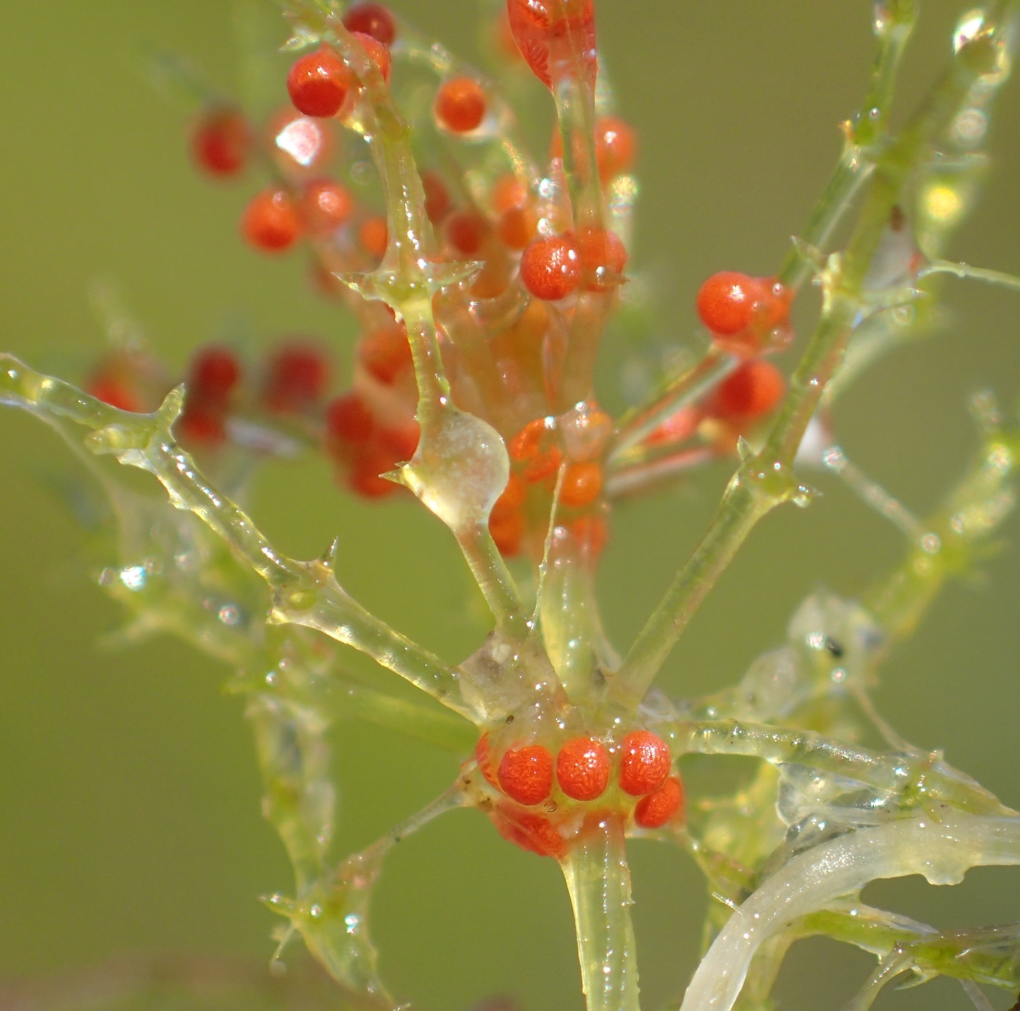
- The male sex organ is spherical, displaying colors ranging from yellow to red, and is known as the globule.
- The female sex organ, called the nucule or oogonium, is oval and typically green. These sex organs develop on the nodes of the branch of limited growth (i.e., primary lateral) and are intermingled with secondary laterals.
The nucule is always situated singly above the globule, creating a distinctive arrangement during sexual reproduction. Most species of Chara are homothallic or monoecious, meaning that male and female sex organs develop on the same plant. However, some species are heterothallic or dioecious, where male and female sex organs are found on different individuals.
Globule Development:
The globule undergoes a well-defined developmental process, originating at the node of branches of limited growth. The development can be described in various stages:
1. A single peripheral cell of each node functions as the antheridial initial. 2. The antheridial initial undergoes a transverse division, forming two cells: the lower one becomes the pedicel cell, forming the stalk, and the upper one becomes the antheridial mother cell.
3. The antheridial mother cell undergoes two vertical divisions at right angles, followed by one transverse division, resulting in an octant (8-celled stage).
4. Each cell of the octant stage undergoes periclinal division, forming the outer 8 and inner 8 cells.
5. Either the outer or inner cells undergo another periclinal division, resulting in 3 layers of 8 cells each.
6. The outer 8 cells form shield cells, the middle 8 cells form the manubrium, and the inner 8 cells form the primary capitula.
7. The primary capitula further divide and form two or more secondary capitula.
8. Each secondary capitulum undergoes division, forming 2-4 antheridial filaments consisting of 25 to 250 antheridial cells, or antheridia, produced through repeated mitotic divisions.

Structure of Mature Globule:
Mature globules are spherical, yellow to red in color, and exhibit a distinctive structure.
- Each globule comprises eight curved plates, known as shield cells, situated towards the outer side.
- From the inner side of each shield cell, a centrally placed rod-shaped structure, called the manubrium, develops.
- At the distal end of each manubrium, one or more globosae cells develop, referred to as the primary capitula.
- Each primary capitulum develops two or more secondary capitula.
- Each secondary capitulum ultimately develops 2–4 long antheridial filaments.
- Each antheridial filament consists of 25–250 cells, with each cell, i.e., antheridium, forming a biflagellate, coiled, and uninucleate antherozoid.
In conclusion, a mature globule can produce as many as 20,000 to 50,000 antherozoids through this intricate developmental process.

Nucule:
Structure and Development: The nucule is oval in shape and contains the central egg, known as the oosphere, surrounded by tube cells that form a corona-like structure on the tapering end.
Development of Nucule: The oogonial initial develops from the peripheral nodal cell or the primary laterals. The oogonial initial undergoes two transverse divisions, forming a 3-celled stage. The lowermost cell is the pedicel cell, the middle one is the nodal cell, and the uppermost one represents the oogonial mother cell. The pedicel cell remains undivided, forming the stalk of the nucule. The middle one undergoes several vertical divisions, forming five sheath initials that surround a central cell. The oogonial mother cell divides transversely, forming a lower stalk cell and an upper egg. The egg elongates further, developing an oval structure with a receptive spot. Large amounts of oil and starch are deposited in the ovum.
The sheath initial elongates further, dividing transversely into upper small cells, the corona cells, which form a crown-like structure at the top of the oogonium. The lower five cells formed are tube cells. The tube cells elongate and become spirally twisted in a clockwise direction outside the oogonium, providing protection to the egg.
Structure of Mature Nucule: The nucule of Chara is oval with a short stalk, developing at the node of the primary laterals just above the globule in homothallic species. It consists of a centrally placed central cell, one stalk, and one large egg at the top. The entire structure is covered from the base by five spirally twisted tube cells, except at the apex, where they form a crown made up of five corona cells.
Process of Fertilization: When the nucule matures, it opens like a flower, and antherozoids move towards it, with only one able to fertilize it. When the antherozoid comes in contact with the egg, it closes, capturing only one antherozoid inside for fertilization. Inside, plasmogamy and karyogamy take place, forming a zygote. Meiosis then occurs, resulting in four nuclei. Only one nucleus becomes active, while the other three denature. The active nucleus divides into two—one moves into the soil, and one remains outside.
Germination: During germination, the nucleus of the oospore migrates towards the upper region, undergoing meiotic division to form four haploid nuclei. The oospore then undergoes germination, leading to the development of a new Chara plant.


.jpg)
0 Comments